Medical & Educational Instruments
Skulls
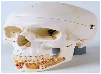
Classic Skull with Opened Lower Jaw, 3-part
In this highly detailed skull, the mandible is opened to show the dental roots with
vessels and nerves. The cranial bones, bone components, fissures, foramina and
other structures are numbered. The cranial sutures are shown in color, as are the
meningeal vessels and venous sinuses. Over 100 features are identified in the
accompanying product manual.
20 x 13.5 x 15.5 cm; 0.7 kg
M-A22
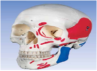
Classic Skull, painted, 3-part
The muscle origins (red) and
insertions (blue) are shown in color
on the left side of the skull. Cranial
bones and structures are numbered
on the right side. The skull
identifies over 140 anatomical
elements.
20 x 13.5 x 15.5 cm; 0.7 kg
M-A23
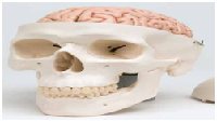
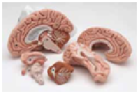
Exceptional detail at an affordable price, the 3-part
Classic Skull with soft 5-part Brain is a first choice
for basic anatomical studies
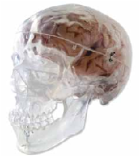
Classic Skull, transparent, 3-part
Use this unique skull to study internal structures that
otherwise are visible only through x-ray images.
• High-quality original cast, hand-made of hard,
unbreakable plastic
• Highly accurate representation of the fissures,
foramina, processes, sutures etc.
• Can be disassembled into skull cap, base of skull and
mandible
• As an option, you can insert a 5-part brain
M-C18 into the skull
20 x 13.5 x 15.5 cm, 0.7 kg
M-A20/T
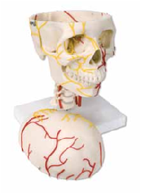
Neurovascular Skull
A life size adult skull with seven cervical vertebrae
mounted upon a stand. The arteries are shown on one
side and nerves on the other. Removing the skull cap
exposes the main nerves and arteries on the floor of
the cranium. The 12 cranial nerves and
the distribution of their branches is also
shown.
29 x 21 x 18.5 cm; 1.3 kg
M-W19018
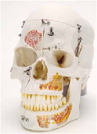
Deluxe Demonstration
Skull, 42-part
This replica of the human
skull has exceptional quality.
The skullcap is removable
and the base of the skull is
midsagitally divided. The frontal
sinus, perpendicular lamina and
vomer are fitted with flaps which can
be opened to view the lateral nose
wall and sphenoidal sinus. On the
left half, the temporal bone can be
removed and folded up in the area
of the tympanic membrane. The
maxilla and mandible are opened
to reveal the alveolar nerves. On the
right side the temporal bone is opened to
reveal the sigmoid sinus, the facial nerve canal and the semicircular
ducts. Additional flaps are located at the maxillary sinus and the right half of
the mandible, so that the dental roots of the premolars and molars of the lower
jaw can also be viewed. The natural occlusion and the individual removal and
replacement of each tooth also make this skull especially interesting for dentists.
• Opened maxilla and mandible to reveal alveolar nerves
• Opened temporal bone reveals sigmoid sinus, facial nerve canal,
and semicircular ducts
• View dental roots of premolars and molars of lower jaw
• Remove and replace all 32 teeth
• Medical quality
• Cast from natural specimen
28 x 22.5 x 18.5 cm; 1.5 kg
M-A27
