Medical & Educational Instruments
Renal System
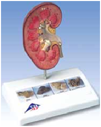
Kidney Stone Model
Here's a great tool to inform patients about kidney stones
(nephrolithiasis) and urinary stones (urolithiasis). It shows
an opened right kidney in natural size. The renal calices,
the renal pelvis, and the ureter are opened as well enabling
concretions or stones to be identified in the following
typical positions:
• In the area of the renal pyramids
• In the area of origin of the upper calix group
• In the renal cortex
• In the connecting tubule of the lower calix group,
causing congestion of the minor calices (partially
closed, partially opened) in the ureter
• 4 original color pictures on the base show various
kidney stones.
14 x 10 x 16.5 cm; 0.18 kg
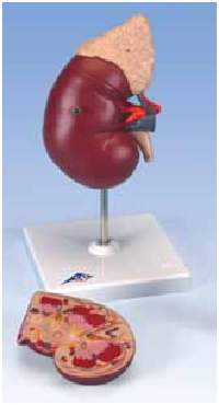
Kidney with Adrenal Gland, 2-part
Affordable and convenient, this model shows:
• Kidney with adrenal gland
• Renal and adrenal vessels
• Upper portion of ureter
The front half of the kidney is removable enabling
demonstration of cortex medulla and vessels as well as
renal pelvis. On stand.
20 x 12 x 12 cm; 0.9 kg
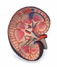
Kidney Section, 3 times full-size
This colorful and anatomically accurate model depicts
a longitudinal section of the human right kidney. All
important structures of the human kidney for student
and patient education are shown. No baseboard
included with the basic kidney section.
8.5 x 19 x 26 cm; 0.9 kg
M-K09
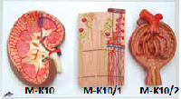
Kidney, Nephrons,Blood Vessels and Renal Corpuscle
A complete series of 3 models mounted on a baseboard
allowing study of the kidney and its different structures
in great detail. Together they show:
• A longitudinal section of the right kidney, 3 times
life-size (M-K10)
• A nephron depicting a section through renal cortex
and medulla. Also features the renal corpuscles with
proximal and distal convoluted tubules, loops of
Henle, collecting tubules, and blood vessels. 120 times
life-size (M-K10/1)
• The third section shows an opened Malpighian
corpuscle with glomerulus and Bowman’s capsule.
700 times full size. (M-K10/2).
29 x 52 x 9 cm; 2.8 kg
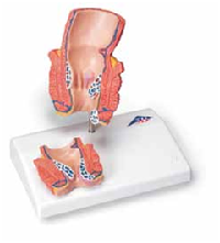
Hemorrhoid Model
A vivid model designed to educate on the subject of
hemorrhoids. The model is a life-size frontal section
of the rectum as well as a smaller relief on a pedestal.
In addition to the anatomical structures of the rectum
(sphincter, mucous membrane, venous plexus), the
model shows internal hemorrhoids during stage I and
II as well as external hemorrhoids. The relief exhibit
shows hemorrhoids during stage III and IV.
Mounted on base.
14 x 10 x 14 cm; 0.2 kg
M-K27
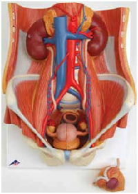
Dual Sex Urinary System, life-size, 6-part
All in one model of the human urinary system. One
front half kidney is removable; easy to change male
insert (bladder and prostate, front and rear half) and
female insert (bladder, womb and ovaries, 2 lateral
halves). On baseboard.
Features include:
• Structures of retroperitoneal cavity
• Large and small pelvis with bones and muscles
• Inferior vena cava
• Aorta with its branches, including iliacal vessels
• Upper urinary tract
• Rectum
• Kidney with adrenal gland.
41 x 31 x 15 cm; 2.3 kg
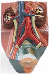
Male Urinary System, ¾ full-size
• Inferior vena cava
• Renal veins
• Aorta with its branches
• Iliacal vessels
• Ureter
• Urinary bladder
• Prostate
• Adrenal gland
• Rectum
• Musculature
The right kidney is opened.
10 x 18 x 26 cm; 1.0 kg
M-VF325