Medical & Educational Instruments
Digestive System
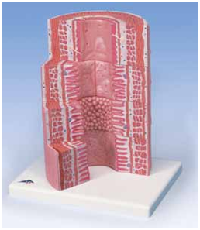
MICROanatomy™ Digestive System
The model illustrates the structure of the
fine tissues of four characteristic sections of
the digestive system:
• Esophagus
• Stomach
• Small intestine
• Large intestine
The front of the model, from top to
bottom, shows a magnified view, in
histological section, of the individual
sections of the digestive system and
their fine tissue structures. The back of
the model has highly magnified views of
didactically interesting areas
of each section shown on the
front.
26 x 29.5 x 18.5 cm; 1.5 kg
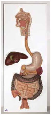
Digestive System
This life-size model demonstrates the path food
takes through the body during digestion. The entire
digestive system is illustrated in graphic relief
featuring:
• Nose • Mouth cavity and Pharynx
• Esophagus • GI tract
• Pancreas • Liver with gall bladder
• Spleen
The duodenum, cecum, and rectum are opened.
The liver, stomach, and transverse colon are
removable. Mounted on baseboard.
81 x 33 x 10 cm; 4.4 kg
2-part model M-K20
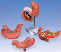
Stomach, 3-part
Shows the layers of the stomach wall from cardia
to pylorus. The front stomach half and the opened
duodenum with pancreas are removable.
Depicted are:
• Layers of stomach wall
• The lower esophagus
• Duodenum
• Pancreas
• Vessels
• Nerves
Delivered on removable stand.
25 x 22 x 12 cm; 0.8 kg
M-K16
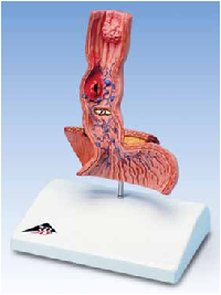
Diseases of the Esophagus
Reaching from the lower part of the
esophagus to the upper part of the
stomach, this vivid, life-size frontal
section shows many common conditions
in this area including:
• Reflux esophagitis
• Ulcer
• Barrett’s Ulcer
• Esophageal carcinoma
• Esophageal varices
• Hiatal hernia
Mounted on base.
14 x 10 x 19 cm; 0.19 kg
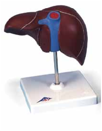
Liver with Gall Bladder
This realistic liver with gall bladder shows:
• 4 lobes with gall bladder
• Extrahepatic ducts
• Hilus vessels.
On removable stand.
18 x 18 x 12 cm; 0.5 kg
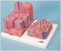
MICROanatomy™ Liver
This 2-part model shows a highly magnified
view of a section of the liver. It illustrates the
functional and structural components of the
liver with two different enlargements. The left
part shows a section of the liver that
comprises several liver lobules. The
right part of the model is a
highly magnified view
of the sectioned liver
lobule on the left.
26 x 15 x 18.5 cm; 0.7 kg
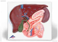
Liver with Gall Bladder,
Pancreas and Duodenum
This highly-detailed relief model
arrives on a baseboard and provides a
detailed look at the liver with:
• Ducts
• Gall bladder
• Pancreas
• Duodenum
• Vessels
• Extra-hepatic ducts with
gall bladder
• Main pancreatic duct and
their orifices
4 x 20 x 18 cm; 0.8 kg
Gallstone Model
The anatomy of the biliary system and its
surroundings are shown at half natural
size in this graphic model for education.
Both acute inflammation (cholecystitis)
and the tissue changes caused by chronic
inflammation can be identified in the
gallbladder wall. Gallstones can be found
in the following typical locations:
• In the fundus area of the gall bladder
• In the area of the spiral valve
• In the area of the common
bile duct
• In the papillary opening to
the small intestine
Mounted on base.
14 x 10 x 19 cm; 0.28 kg
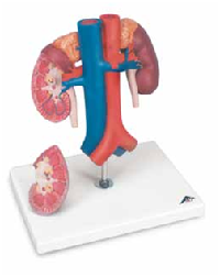
Kidneys with Vessels, 2-part
Life size model presenting the kidneys with suprarenal
glands, the outgoing ureters, the renal vessels, and the
large vessels situated close to the kidneys. The front half
of the right kidney can be removed to reveal the renal
pelvis, the renal calices, the renal cortex, and the renal
medulla. Comes on stand.
21 x 18 x 28 cm; 1.0 kg
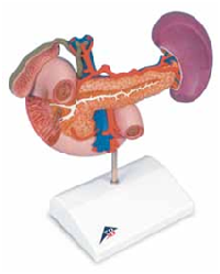
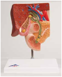
Rear Organs of Upper Abdomen
Representing the duodenum, which is partially opened,
along with opened gall bladder and bile ducts. The
model also includes the pancreas, revealing large ducts,
the spleen, and the surrounding vessels, all in natural
size. Comes on stand.
23 x 12 x 20 cm; 0.55 kg
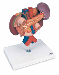
Kidneys with Rear Organs
of Upper Abdomen, 3-part
A combination of models M-K22/1 and M-K22/2. The
upper abdominal organs are attached in their natural
positions and are removable from the kidneys.
Comes on stand.
24 x 18 x 29 cm; 1.4 kg
M-K22/3